
Comparative Evaluation of HER2 Overexpression in Breast Carcinoma Using Cell Blocks and Corresponding Formalin-Fixed Paraffin-Embedded Tissue Blocks: A Prospective Study
Main Article Content
Keywords
Breast carcinoma, HER-2 overexpression, Cell block, Histological tissue block, Immunohistochemistry
Abstract
Background: Breast cancer is one of the most common cancers among women in Nigeria. Human epidermal growth factor receptor 2 (HER2) is an important prognostic and predictive biomarker that guides targeted therapy. The tumour grade is an important prognostic factor and is also important in the treatment of patients. In a resource-limited setting, cell block cytology may serve as an alternative for initial biomarker assessment and also as an initial diagnostic tool for planning definitive management. The study aims to compare HER2 overexpression of breast carcinoma using cell blocks and corresponding paraffin wax-embedded (FFPE) tissue blocks and to evaluate the concordance between both methods.
Methodology: This was a one-year prospective study involving 83 cases of breast carcinoma patients with both cell block and corresponding FFPE tissue specimens. HER2 immunohistochemistry was performed using the ASCO/CAP 2018 guideline. Sensitivity, specificity, positive predictive value (PPV), and negative predictive value (NPV) were calculated with 95% confidence intervals (CI). Concordance was assessed using Cohen’s kappa statistic. McNemar’s test was used for paired comparisons.
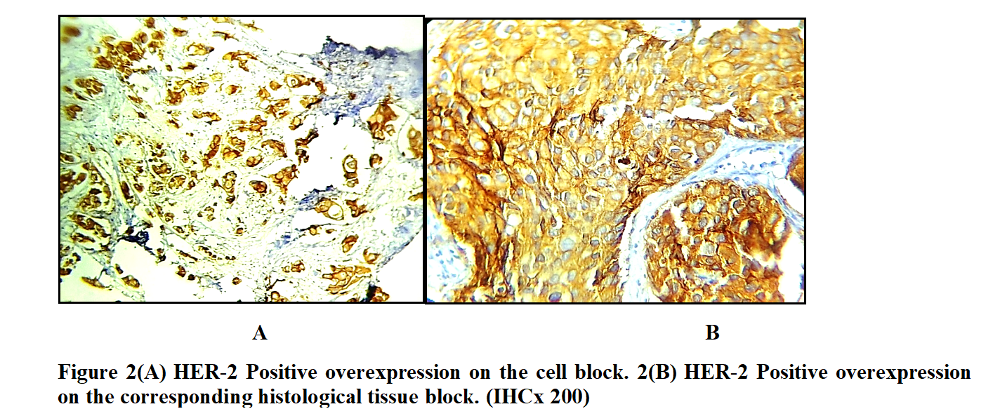
Results: The mean age of the study participants was 43.1 ±13.1 years, with a peak age group of 40-49 years. IHC HER2 overexpression was done on both cell blocks and histological blocks. In cell blocks, HER2 expression showed 15 cases (18.1%), 65 cases (78.3%), 3 cases (3.6%) were positive, negative, and equivocal, respectively while from histologic tissues, 15 cases (18.1%), 63 cases (75.9%), 5 cases (6.0%) were also positive, negative and equivocal respectively. The overall concordance rate between the two methods was 93.5%, with concordance rates of 100% for HER2-positive cases, 96.9% for HER2-negative cases, and 60% for equivocal cases. Sensitivity and specificity of cell block HER2 assessment were 96.9% (95% CI: 82.9-99.9) and 100% (95% CI: 94.3-100.0), respectively. The PPV of HER2 assessment on cell block was 100.0% (95% CI: 78.2-100.0), and the NPV was 97.1% (95% CI: 89.9-99.6). The kappa coefficient for agreement was 0.935, indicating excellent agreement. McNemar’s test showed no statistically significant difference (p = 0.480). Equivocal (2+) cases were included without FISH confirmation. Most of the cases were invasive ductal carcinoma (NST), accounting for 97.6% (81 cases).
Conclusion: Cell block cytology demonstrates strong concordance with FFPE tissue for HER2 assessment and may serve as a reliable alternative for initial triaging in resource-limited settings, particularly where tissue is not readily feasible. However, confirmatory testing on tissue biopsy remains essential, particularly for equivocal cases.
References
Sung H, Ferlay J, Siegel RL, Laversanne M, Soerjomataram I, Jemal A, et al. Global Cancer Statistics 2020: GLOBOCAN Estimates of Incidence and Mortality Worldwide for 36 Cancers in 185 Countries. CA Cancer J Clin. 2021;71:209–249.
Joko-Fru WY, Jedy-Agba E, Korir A, Ogunbiyi O, Dzamalala CP, Chokunonga E, et al. The evolving epidemic of breast cancer in sub-Saharan Africa: results from the African Cancer Registry Network. Int J Cancer. 2020;147:2131-2141.
Allemani C, Matsuda T, Di Carlo V, Harewood R, Matz M, Niksic M, et al. Global surveillance of trends in cancer survival 2000-14 (CONCORD-3): analysis of individual records for 37 513 025 patients diagnosed with one of 18 cancers from 322 population-based registries in 71 countries. Lancet. 2018;391:1023-1075.
Yusuf I, Atanda AT, Umar AB, Imam MI, Mohammed AZ, Ochicha O, et al. Cancer in Kano, Northwestern Nigeria: A 10-year update of the Kano cancer registry. Ann Trop Pathol. 2017;8:87-93.
Tsoka-Gwegwenia JM, Cumber SN, Nchanji KN. Breast cancer among women in sub-Saharan Africa: prevalence and a situational analysis. South Afr J Gynaecol Oncol. 2017;9(2):28-30.
Ibrahim IM, Iliyasu Y, Mohammed AZ. Histopathological review of breast tumours in Kano, Northern Nigeria. Sub-Saharan Afr J Med. 2015;2:47-51.
Stanley N, Anyanwu C. Breast cancer in Eastern Nigeria: A ten-year review. West Afr Med J. 2000;21:120-125.
Oguntunde PE, Adejumo AO, Okagbue HI. Breast cancer patients in Nigeria: data exploration approach. Data Brief. 2017;15:47-57.
Dauda AM, Misauno MA, Ojo EO. Histopathological types of breast cancer in Gombe, North Eastern Nigeria: A seven-year review. Afr J Reprod Health. 2011;15(1):107-109.
Kene TS, Odigie VI, Yusufu LM, Yusuf BO, Shehu SM, Kase JT. Pattern of presentation and survival of breast cancer in a teaching hospital in North Western Nigeria. Oman Med J. 2010;25(2):104-107.
Ezeome ER, Emegoakor CD, Chianakwana GU, Anyanwu SNC. The pattern of male breast cancer in Eastern Nigeria: A 12-year review. Nig Med J. 2010;51(1):26-29.
Amir H, Kaaya EE, Kwesigabo G, Kiitinya JN. Breast cancer before and during the AIDS epidemic in women and men: a study of Tanzanian Cancer Registry Data 1968 to 1996. J Natl Med Assoc. 2000;92:301-305.
Othieno-Abinya NA, Nyabola LO, Abwao HO, Ndege P. Postsurgical management of patients with breast cancer at Kenyatta National Hospital. East Afr Med J. 2002;79:156-162.
Muller K, Jorns JM, Tozbikian G. What’s new in breast pathology 2022: WHO 5th edition and biomarker updates. J Pathol Transl Med. 2022;56:170-171.
Adeniji KA, Bello JM, Durowade KA. Rising pattern of breast cancer in young women. East Afr Med J. 2015;92(6):279-283.
Omoniyi-Esan GO, Osasan S, Titiloye N, Olasode B. Cytopathological review of breast lesions in Ile-Ife, Nigeria. Internet J Third World Med. 2008;8:106-112.
Ngadda HA, Gali BM, Bakari AA, Yawe-Terna EH, Tahir MB, Apari E, et al. The spectrum of female breast diseases among the Nigerian population in the Sahel climatic zone. J Med Med Sci. 2011;2:1157-1161.
Boder JE, Elmabrouk Abdalla FB, Elfageih MA, Abusaa A, Buhmeida A, Collan Y. Breast cancer patients in Libya: Comparison with European and central African patients. Oncol Lett. 2011;2:323-330.
Awadelkarim KD, Arizzi C, Elamin EO, Hamad HM, De Blasio P, Mekki SO, et al. Pathological, clinical, and prognostic characteristics of breast cancer in Central Sudan versus Northern Italy: implications for breast cancer in Africa. Histopathology. 2008;52(4):445-456.
Duran MC, Vega F, Bueno GM, et al. Characterization of tumoral markers correlated with ErbB2 (Her2/Neu) over-expression and metastasis in breast cancer. Proteomics Clin Appl. 2008;2:1313-1326.
Wolff AC, Hammond MEH, Allison KH, Harvey BE, Mangu PB, Bartlett JMS, et al. Human Epidermal Growth Factor Receptor 2 Testing in Breast Cancer: American Society of Clinical Oncology/College of American Pathologists clinical practice guideline focused update. J Clin Oncol. 2018;36(20):2105-2122.
Bansal M, Bagga P, Singh S, Paul S. Comparative evaluation of immunohistochemical expression of estrogen receptor, progesterone receptor, and HER2 in fine needle aspiration cell blocks and surgical biopsies in primary breast carcinoma. Int J Contemp Med Res. 2020;7.
Kumar SK, Gupta N, Rajwanshi A, Joshi K, Singh G. Immunochemistry for oestrogen receptor, progesterone receptor and HER2 on cell blocks in primary breast carcinoma. Cytopathology. 2012;23:181-186.
Vohra P, Buelow B, Chen YY, Serrano M, Vohra MS, Berry A, et al. Estrogen receptor, progesterone receptor, and human epidermal growth factor receptor 2 expression in breast cancer FNA cell blocks and paired histologic specimens: A large retrospective study. Cancer Cytopathol. 2016;124:828-835.
Nishimura R, Okamoto N, Tanaka S. HER2 immunohistochemistry for breast cancer cell blocks can be used in the same way as that used for histological specimens. Diagn Cytopathol. 2016;44:274-279.
Dong J, Ly A, Arpin R, Ahmed Q, Brachtel E. Breast fine needle aspiration continues to be relevant in a large academic medical centre: experience from Massachusetts General Hospital. Breast Cancer Res Treat. 2016;158:297-305.
Navani S, Bhaduri AS. High incidence of oestrogen receptor negative progesterone receptor positive phenotype in Indian breast cancer: fact or fiction? Indian J Pathol Microbiol. 2005;48:199-201.
Rhodes A, Jasani B, Balaton AJ, Barnes DM, Anderson E, Bobrow LG, et al. Study of interlaboratory reliability and reproducibility of estrogen and progesterone receptor assays in Europe. Documentation of poor reliability and identification of insufficient microwave antigen retrieval time as a major contributory element of unreliable assays. Am J Clin Pathol. 2001;115:44-58.
Beatty BG, Bryant R, Wang W, Ashikaga T, Gibson PC, Leiman G, et al. HER-2/neu detection in fine-needle aspirates of breast cancer: fluorescence in situ hybridization and immunocytochemical analysis. Am J Clin Pathol. 2004;122:246-255.
Nizzoli R, Bozzetti C, Crafa P, Naldi N, Guazzi A, Di Blasio B, et al. Immunocytochemical evaluation of HER-2/neu on fine-needle aspirates from primary breast carcinomas. Diagn Cytopathol. 2003;28:142-146.
Article Sidebar
Article Details

This work is licensed under a Creative Commons Attribution-NonCommercial-ShareAlike 4.0 International License.
This is an open-access journal and articles are distributed under the terms of the Creative Commons Attribution Non-Commercial Share-Alike License 4.0. This licence allows users to download and share, remix, tweak and build upon the article for non-commercial purposes, so long as the original authorship is acknowledged and the new creations are licensed under identical terms.
Adeyemi Sofoluwe Gbenga, Department of Anatomic Pathology, Federal University of Health Sciences Azare (FUHSA), Bauchi State, Nigeria
Department of Histopathology, Federal University of Health Sciences Teaching Hospital, Azare (FUHSTHA), Bauchi State
Rufai Yunusa, Department of Anatomic Pathology, Federal University of Health Sciences Azare (FUHSA), Bauchi State, Nigeria
Department of Histopathology, Federal University of Health Sciences Teaching Hospital, Azare (FUHSTHA), Bauchi State
Ahmad Hamza, Department of Anatomic Pathology, Federal University of Health Sciences Azare (FUHSA), Bauchi State, Nigeria
Department of Histopathology, Federal University of Health Sciences Teaching Hospital, Azare (FUHSTHA), Bauchi State.
Taoheed Atanda Akinfenwa, Department of Histopathology, Aminu Kano Teaching Hospital, Kano, Kano State
Department of Anatomical Pathology, Faculty of Basic Clinical Sciences, Bayero University Kano, Kano State
Yusuf Ibrahim , Department of Histopathology, Aminu Kano Teaching Hospital, Kano, Kano State
Department of Anatomical Pathology, Faculty of Basic Clinical Sciences, Bayero University Kano, Kano State
Simon Ezenkwa Uchenna, Department of Anatomic Pathology, Federal University of Health Sciences Azare (FUHSA), Bauchi State, Nigeria
Department of Histopathology, Federal University of Health Sciences Teaching Hospital, Azare (FUHSTHA), Bauchi State
Ajanaku Jimoh Abdulrazaq, Department of Anatomic Pathology, Federal University of Health Sciences Azare (FUHSA), Bauchi State, Nigeria
Department of Histopathology, Federal University of Health Sciences Teaching Hospital, Azare (FUHSTHA), Bauchi State.